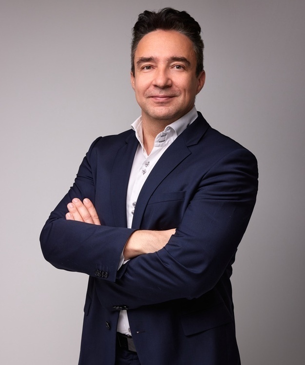
Special Seminar with Dr. Balázs Rózsa: Real-Time 3D Imaging and Photostimulation in Freely Moving Animals: A Novel Approach Using Robotic Acousto-Optical Microscopy
Description
Speaker: Dr. Balázs Rózsa
Director, BrainVisionCenter Research Institute; Head of the Laboratory of 3D functional network and dendritic imaging, HUN-REN IEM; Head of the Laboratory of Neural Circuits and Computation, PPCU; Founder, Femtonics Ltd.
Abstract:
Our long-term goal is to explore the feasibility of creating a visual prosthetic using direct 3D cortical photostimulation. To achieve this aim, developing a robust and reliable behavioral protocol is just as crucial as advancing imaging technology. Current solutions either offer excellent optical quality but limit animal motion, causing significant stress that disturbs behavioral results and reliability, or they allow free movement but with limited optical quality. Beyond visual prosthetics development, there is a growing demand from researchers, pharmaceutical companies, and biotech firms to test their pharmacological and gene therapy products, as well as innovative therapeutic and diagnostic tools, in freely moving animals. While several advanced head-mounted microscopes with one-, two-, and even three-photon excitation (such as light field microscopy, Mini2p-Scope, and Inscopix's Miniscope) offer good imaging capabilities in freely moving animals, they face challenges due to their small scanners and the limited number and diameter of lenses in their objectives, compared to full-sized microscopes, which are too heavy for animals to carry. These limitations result in smaller fields of view (FOV), lower numerical aperture (NA), reduced resolution and detection efficiency, limited fast z-scanning range, and slower scanning speed.
3D acousto-optical (AO) microscopy can address these technical challenges by increasing the product of detection efficiency and measurement speed by six orders of magnitude. However, conventional 3D AO microscopes weigh half a ton, making them impractical for use with freely moving animals. In this talk we will present a unique 3D AO microscope featuring a flexible objective arm with six plus one degrees of freedom, moved by a 6D robotic arm. This system can track freely moving animals with minimal force (1-10 mN) corresponding to only 0.1-1 gram. Our fast, closed-loop, real-time motion correction method, based on local intelligence, effectively eliminates optical errors caused by the deformation of the long objective arm during motion, as well as motion artifacts resulting from breathing, heartbeat, and physical movement of the mice. This cost-effective solution enables high-speed (100 kHz/ROI), real-time, motion-corrected 3D imaging with subcellular resolution, across large scanning volumes. This allows for unique 3D measurement modes such as accelerated learning and behavioral experiments, as well as simultaneous fast 3D voltage imaging and photostimulation of somata, dendrites and spines in full cortical columns in freely moving configurations. By combining the advantages of high-quality imaging with unrestricted animal movement, our system offers a promising approach for advancing visual prosthetics research and meeting the growing demands of the biomedical research community.

