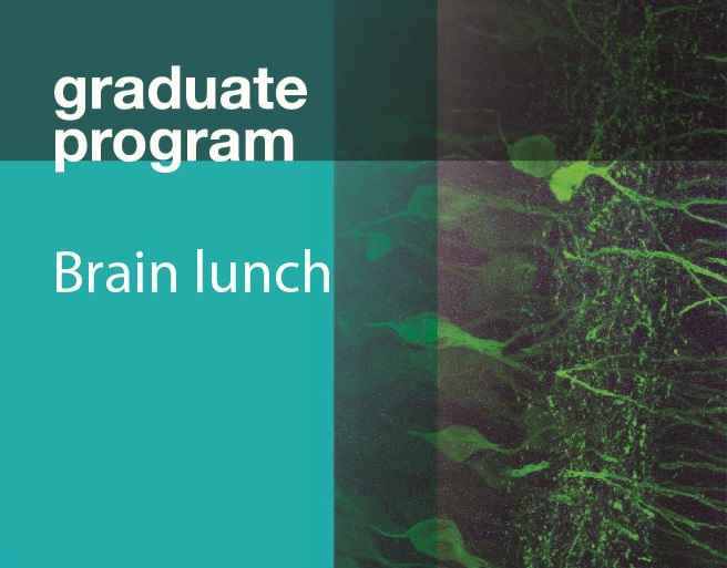
Multiscale proteomic imaging of the brain
Description
The biology of multicellular organisms is coordinated across multiple size scales, from the subnanoscale of molecules to the macroscale, tissue-wide interconnectivity of cell populations. I will introduce a method for super-resolution imaging of the multiscale organization of intact tissues. The method, called magnified analysis of the proteome (MAP), linearly expands entire brain fourfold while preserving their overall architecture and 3D proteome organization. MAP is based on the observation that preventing crosslinking within and between endogenous proteins during hydrogel-tissue hybridization allows for natural expansion upon protein denaturation and dissociation. The expanded tissue preserves its protein content, its fine subcellular details, and its organ-scale intercellular connectivity. Off-the-shelf antibodies are used for multiple rounds of immunolabeling and imaging of a tissue’s magnified proteome. Specimen size can be reversibly modulated to image both inter-regional connections and fine synaptic architectures in the mouse brain.
UPCOMING BRAIN LUNCH TALKS
- 11/21/16 - Naomi Habib, Zhang Lab
- 11/28/16 - Or Shemesh, Boyden Lab
- 12/5/16 - Teruhiro Okuyama, Tonegawa Lab

