Dopamine signals when a fear can be forgotten

Dangers come but dangers also go, and when they do, the brain has an “all-clear” signal that teaches it to extinguish its fear. A new study in mice by MIT neuroscientists shows that the signal is the release of dopamine along a specific interregional brain circuit. The research therefore pinpoints a potentially critical mechanism of mental health, restoring calm when it works, but prolonging anxiety or even post-traumatic stress disorder when it doesn’t.
“Dopamine is essential to initiate fear extinction,” says Michele Pignatelli di Spinazzola, co-author of the new study from the lab of senior author Susumu Tonegawa, Picower Professor of biology and neuroscience at the RIKEN-MIT Laboratory for Neural Circuit Genetics within The Picower Institute for Learning and Memory at MIT, and a Howard Hughes Medical Institute (HHMI) investigator.
In 2020, Tonegawa’s lab showed that learning to be afraid, and then learning when that’s no longer necessary, result from a competition between populations of cells in the brain’s amygdala region. When a mouse learns that a place is “dangerous” (because it gets a little foot shock there), the fear memory is encoded by neurons in the anterior of the basolateral amygdala (aBLA) that express the gene Rspo2. When the mouse then learns that a place is no longer associated with danger (because they wait there and the zap doesn’t recur), neurons in the posterior basolateral amygdala (pBLA) that express the gene Ppp1r1b encode a new fear extinction memory that overcomes the original dread. Notably, those same neurons encode feelings of reward, helping to explain why it feels so good when we realize that an expected danger has dwindled.
In the new study, the lab, led by former members Xiangyu Zhang and Katelyn Flick, sought to determine what prompts these amygdala neurons to encode these memories. The rigorous set of experiments the team reports in the Proceedings of the National Academy of Sciences show that it’s dopamine sent to the different amygdala populations from distinct groups of neurons in the ventral tegmental area (VTA).
“Our study uncovers a precise mechanism by which dopamine helps the brain unlearn fear,” says Zhang, who also led the 2020 study and is now a senior associate at Orbimed, a health care investment firm. “We found that dopamine activates specific amygdala neurons tied to reward, which in turn drive fear extinction. We now see that unlearning fear isn’t just about suppressing it — it’s a positive learning process powered by the brain’s reward machinery. This opens up new avenues for understanding and potentially treating fear-related disorders, like PTSD.”
Forgetting fear
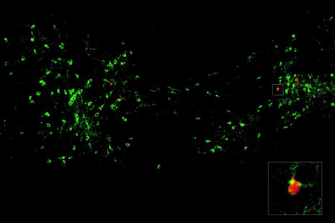
The VTA was the lab’s prime suspect to be the source of the signal because the region is well known for encoding surprising experiences and instructing the brain, with dopamine, to learn from them. The first set of experiments in the paper used multiple methods for tracing neural circuits to see whether and how cells in the VTA and the amygdala connect. They found a clear pattern: Rspo2 neurons were targeted by dopaminergic neurons in the anterior and left and right sides of the VTA. Ppp1r1b neurons received dopaminergic input from neurons in the center and posterior sections of the VTA. The density of connections was greater on the Ppp1r1b neurons than for the Rspo2 ones.
The circuit tracing showed that dopamine is available to amygdala neurons that encode fear and its extinction, but do those neurons care about dopamine? The team showed that indeed they express “D1” receptors for the neuromodulator. Commensurate with the degree of dopamine connectivity, Ppp1r1b cells had more receptors than Rspo2 neurons.
Dopamine does a lot of things, so the next question was whether its activity in the amygdala actually correlated with fear encoding and extinction. Using a method to track and visualize it in the brain, the team watched dopamine in the amygdala as mice underwent a three-day experiment. On Day One, they went to an enclosure where they experienced three mild shocks on the feet. On Day Two, they went back to the enclosure for 45 minutes, where they didn’t experience any new shocks — at first, the mice froze in anticipation of a shock, but then relaxed after about 15 minutes. On Day Three they returned again to test whether they had indeed extinguished the fear they showed at the beginning of Day Two.
The dopamine activity tracking revealed that during the shocks on Day One, Rspo2 neurons had the larger response to dopamine, but in the early moments of Day Two, when the anticipated shocks didn’t come and the mice eased up on freezing, the Ppp1r1b neurons showed the stronger dopamine activity. More strikingly, the mice that learned to extinguish their fear most strongly also showed the greatest dopamine signal at those neurons.
Causal connections
The final sets of experiments sought to show that dopamine is not just available and associated with fear encoding and extinction, but also actually causes them. In one set, they turned to optogenetics, a technology that enables scientists to activate or quiet neurons with different colors of light. Sure enough, when they quieted VTA dopaminergic inputs in the pBLA, doing so impaired fear extinction. When they activated those inputs, it accelerated fear extinction. The researchers were surprised that when they activated VTA dopaminergic inputs into the aBLA they could reinstate fear even without any new foot shocks, impairing fear extinction.
The other way they confirmed a causal role for dopamine in fear encoding and extinction was to manipulate the amygdala neurons’ dopamine receptors. In Ppp1r1b neurons, over-expressing dopamine receptors impaired fear recall and promoted extinction, whereas knocking the receptors down impaired fear extinction. Meanwhile in the Rspo2 cells, knocking down receptors reduced the freezing behavior.
“We showed that fear extinction requires VTA dopaminergic activity in the pBLA Ppp1r1b neurons by using optogenetic inhibition of VTA terminals and cell-type-specific knockdown of D1 receptors in these neurons,” the authors wrote.
The scientists are careful in the study to note that while they’ve identified the “teaching signal” for fear extinction learning, the broader phenomenon of fear extinction occurs brainwide, rather than in just this single circuit.
But the circuit seems to be a key node to consider as drug developers and psychiatrists work to combat anxiety and PTSD, Pignatelli di Spinazzola says.
“Fear learning and fear extinction provide a strong framework to study generalized anxiety and PTSD,” he says. “Our study investigates the underlying mechanisms suggesting multiple targets for a translational approach, such as pBLA and use of dopaminergic modulation.”
Marianna Rizzo is also a co-author of the study. Support for the research came from the RIKEN Center for Brain Science, the HHMI, the Freedom Together Foundation, and The Picower Institute.

